
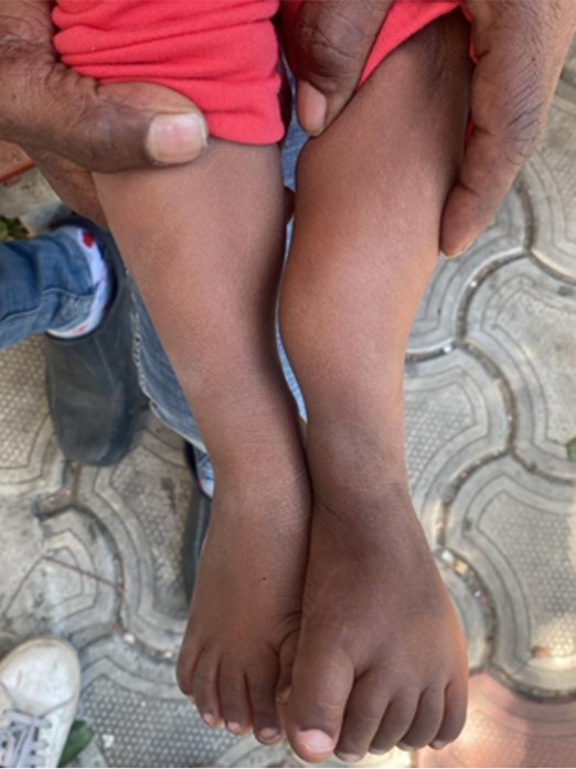
A 2 years old male child presented with progressive bowing of left leg.
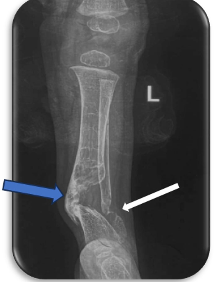
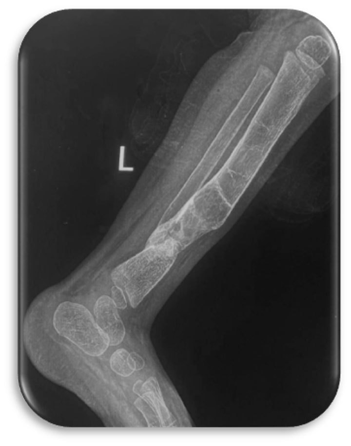
Radiograph of left leg shows abnormal tibial bowing with a bony defect in distal tibia (blue arrow), with cupping of proximal bony fragment and pencil like tapering of distal fragment giving appearance of a joint. Similar defect was also seen in fibula (white arrow).

On MRI, this bony defect is hypointense on T1W and hyperintense on T2W imaging
Based on imaging findings a diagnosis of tibial and fibular pseudoarthrosis was made
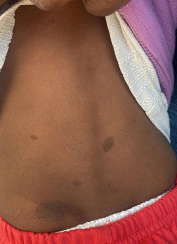
Following the radiologic diagnosis, a careful clinical examination was performed. Multiple café-au-lait macules were identified on the trunk
Putting the pieces together led to final diagnosis: Neurofibromatosis type I (NF-1) with pseudoarthrosis of tibia and fibula
Learning points:
Pseudoarthrosis of tibia: A rare but classic musculoskeletal manifestation of NF1.
Radiology and clinical examination go hand in hand. Don’t only interpret the images- examine the patient.
Pseudoarthrosis of the tibia in a young child should always prompt a search for stigmata of NF1.