
A 33 young male patient presented with progressive weakness of bilateral lower limbs and truncal weakness for 4 years.
He also had a palpable swelling in the posterior aspect of the right thigh.
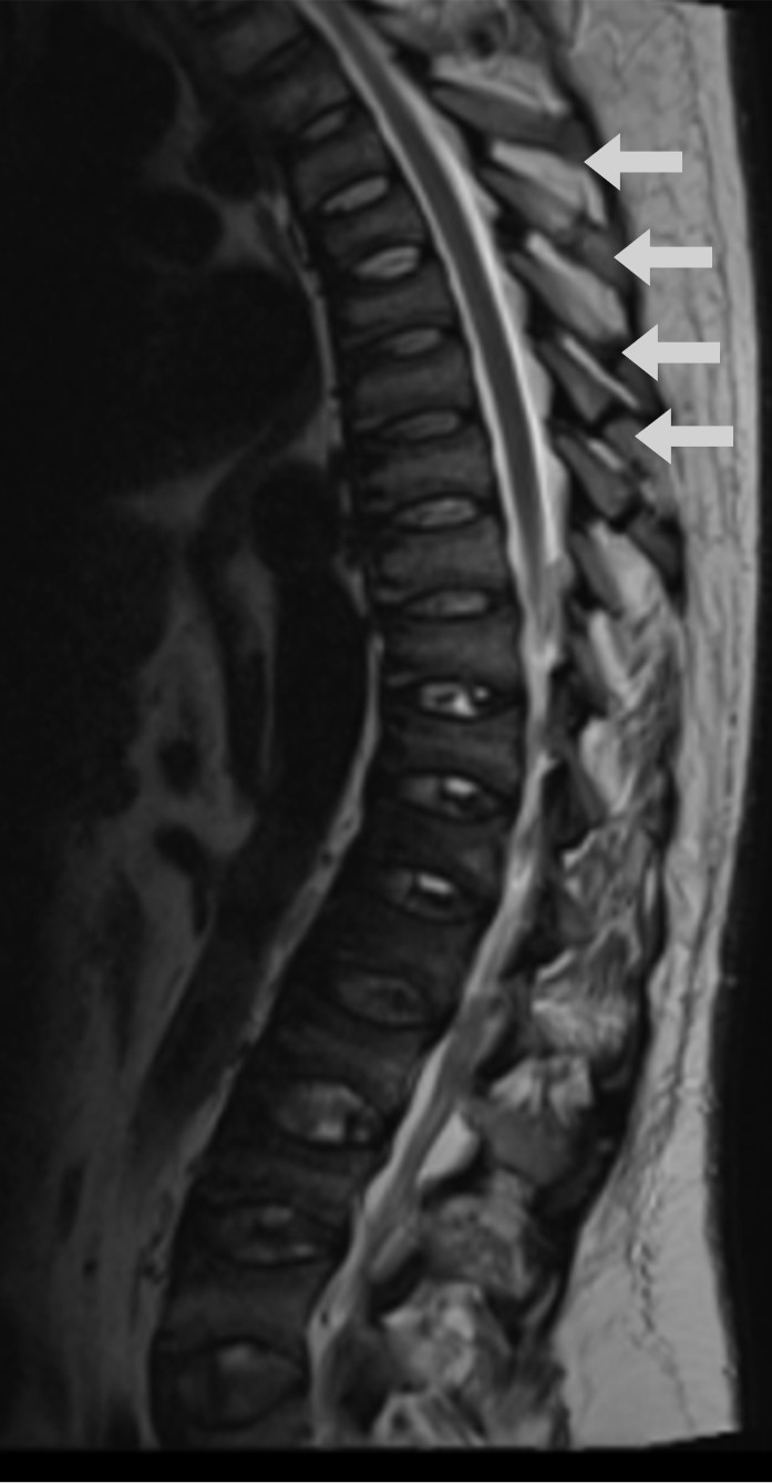
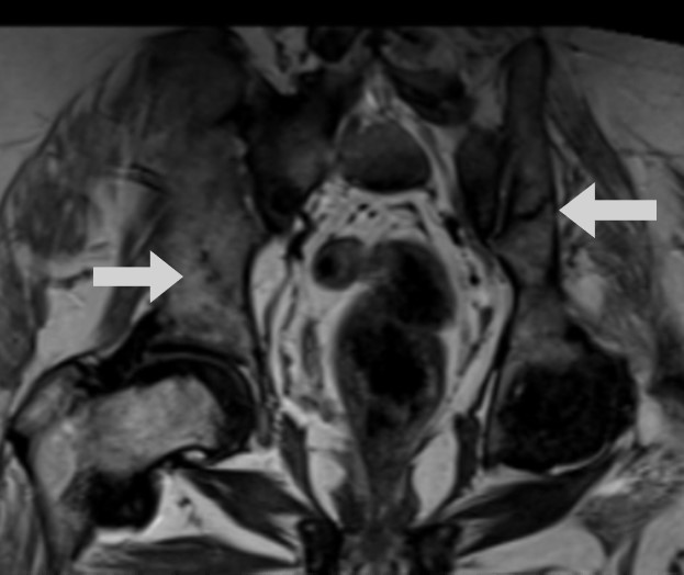
MRI of the spine and pelvis showed multiple insuffiiciency fractures involving the spinous processes of multiple vertebrae and bilateral ilium (arrows).
Multilevel central compression of vertebral bodies were also noted giving a biconcave shaped vertebra (codfish vertebra)
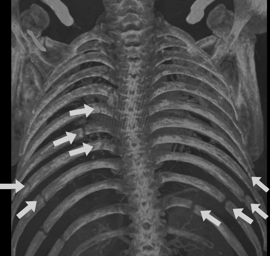
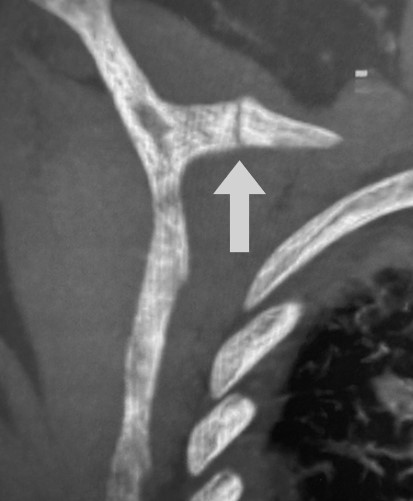
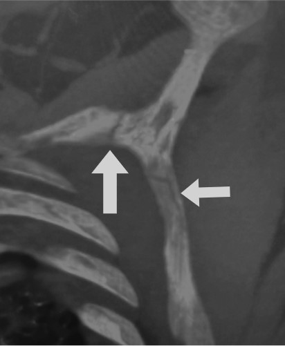
Maximum Intensity projection (MIP) image of bone window of the CT chest also showed multiple insufficiency fractures/ Looser zones involving bilateral ribs and scapulae in addition to the spine (arrows) suggesting severe osteomalacia.
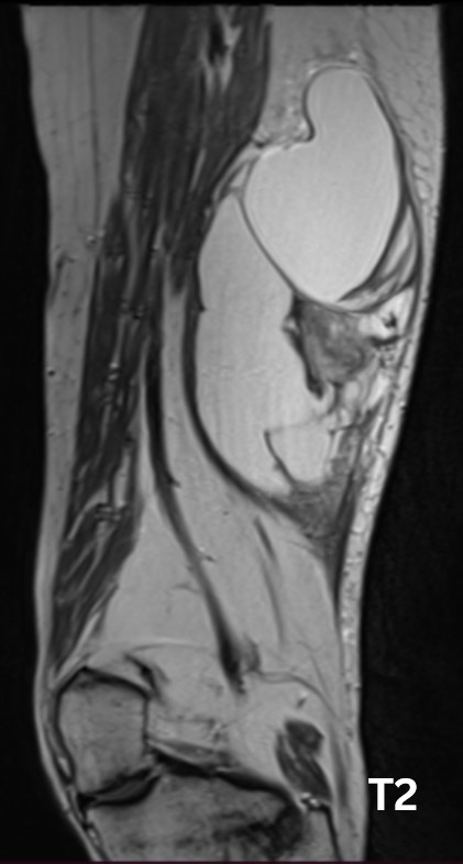
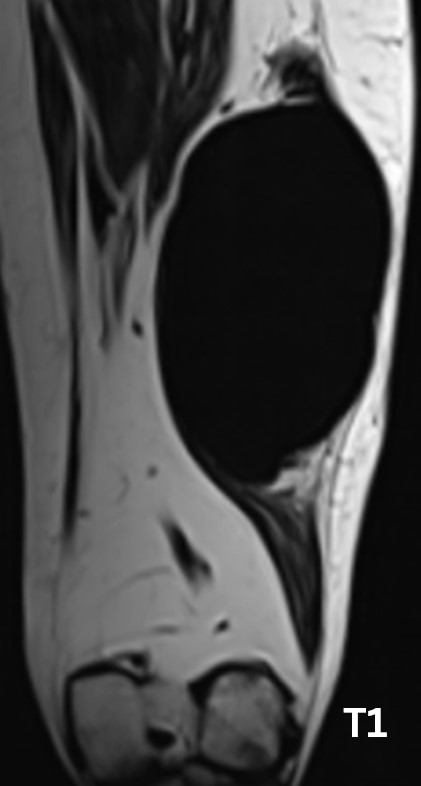
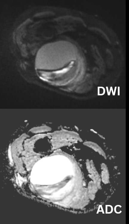
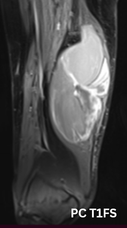
Contrast enhanced MRI of the right thigh was done in view of the palpable swelling, which showed a solid-cystic intermuscular lesion in the posterior compartment of mid thigh. Multiple septations and nodular solid areas which showed diffuse restriction and avid enhancement were noted within.
A diagnosis of phosphaturic mesenchymal tumor with tumor-induced osteomalacia was made and biochemical correlation was suggested.
Biochemical evaluation showed hypophosphatemia, phosphaturia and low vitamin D levels.
FGF-23 levels were significantly high (>26000 pg/ml).
DOTA-PET also showed DOTA avidity within the lesion in the right thigh.
Patient underwent wide local excision of the lesion and post operative radiotherapy with Calcium and Vit D supplementation.
Final HPE of lesion – Low grade fibromyxoid sarcoma.
On follow up, his biochemical parameters including FGF-23 levels normalised with improvement in clinical symptoms
LEARNING POINTS:
- Tumor-Induced osteomalaciais a paraneoplastic syndrome where tumors produce FGF-23.
- Symptoms are often non-specific, making diagnosis difficult.
- Biochemical features include hypophosphatemia, low Vit D3 levels and increased FGF-23 levels.
- Phosphaturicmesenchymal tumors may arise from bone/soft tissue/ skin and imaging features may vary accordingly.
- FN1-FGFR1 and FN1-FGF1 fusion gene are seen upto50% of these tumors.
- 68Ga-DOTA-PET is highly sensitive especially in diagnosing small/ non-palpable tumors.