

50 y male with acute onset pain, redness and swelling over the left shoulder

Plain Radiographs
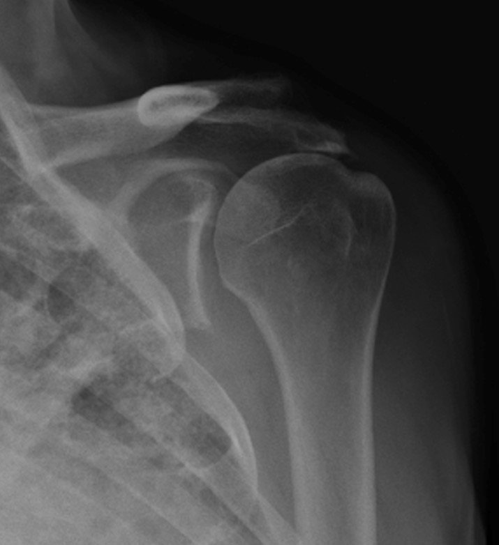
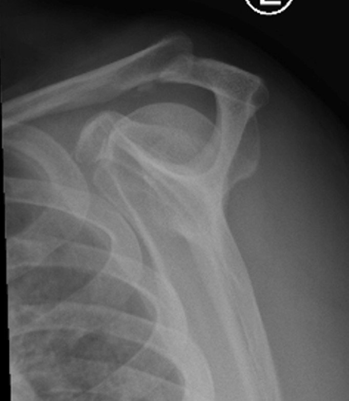
Anteroposterior and Scapular-Y views of the left shoulder at presentation showed grossly reduced acromioclavicular distance and generalised osteopenia. No obvious bone erosions/ focal lesions seen.
MRI Left shoulder
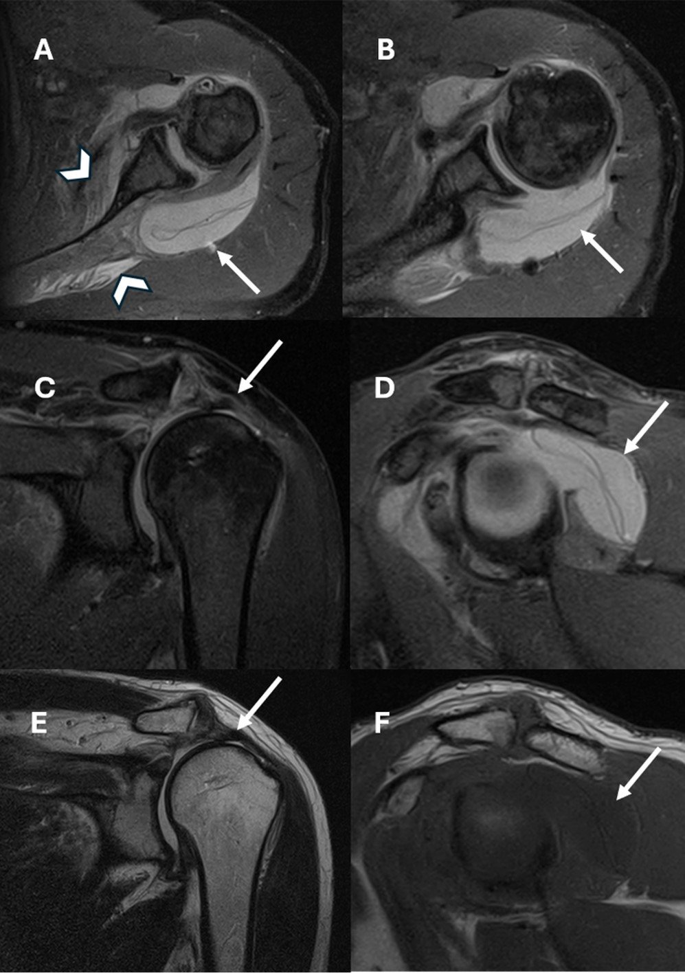
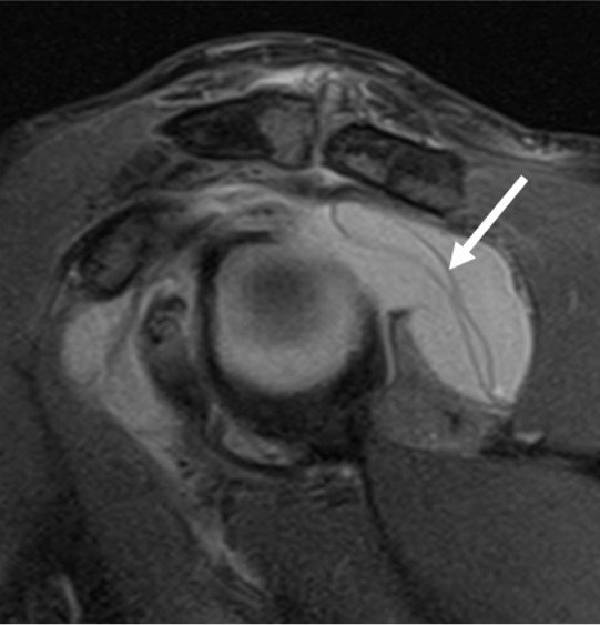
MRI revealed signs of rotator cuff tear with superiorly subluxed humeral head. In addition, there is gross joint effusion with biceps tenosynovitis, and muscle edema within the torn rotator cuff muscles (arrow heads in Image A).
All these point towards INFECTION!!!
Wait! WHAT IS THIS??
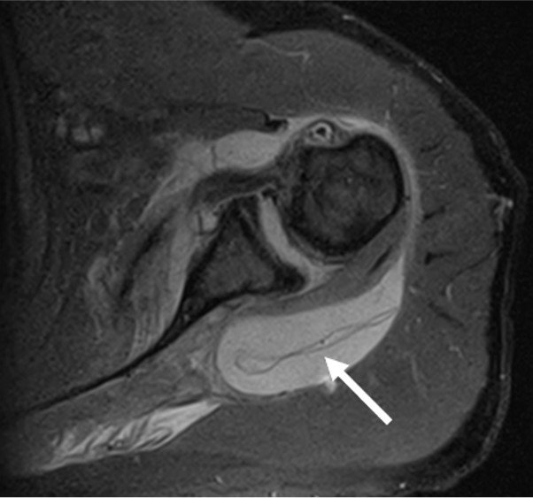
Patient’s previous Imaging were reviewed.
Patient had underwent radiographs and MRI of Left shoulder 1 month before this episode
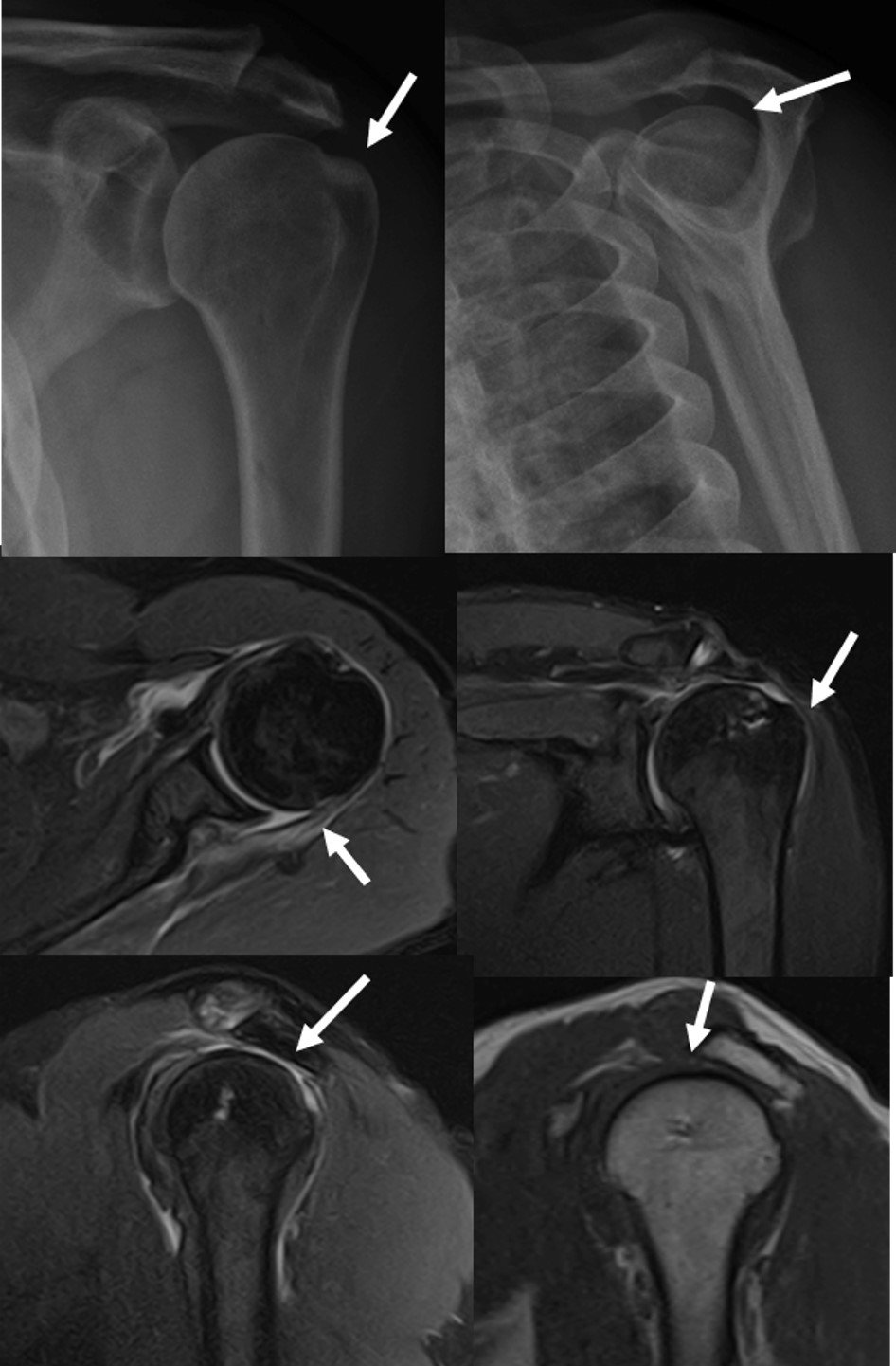
The rotator cuff tear was similar but there were no signs of infection in the previous MRI.
So, what happened in this 1 month?
Patient had undergone Left shoulder arthroscopic sub-acromial space decompression and INspace balloon placement with acromioplasty 3 weeks ago.
Post procedure, with less than satisfactory outcomes with couple of fever spikes and elevated CRP (58 mg/L)
Spacer had ruptured, migrated and got infected.
Arthroscopic removal of infected subacromial in-space balloon and soft tissue debridement, irrigation and lavage were performed.
Analysis and culture showed high granulocytes with Negative gram stain / aerobic or anerobic culture.
Subacromial balloon spacer
- It is a device used in the treatment of patients with massive, irreparable rotator cuff tears.
- It is inserted between the acromion and the humeral head by arthroscopy and filled with saline. The underlying principle – restore the glenohumeral joint congruency and allow frictionless movement and joint rehabilitatiob.
- A less invasive alternative to reverse shoulder arthroplasty with good outcomes.
- 3-17 % complication rate
- Most common complication is balloon rupture, migration and infection, necessitating removal
- This patient went on to have a reverse total shoulder arthroplasty.
(Wright MA, Abboud JA, Murthi AM. Subacromial Balloon Spacer Implantation. Curr Rev Musculoskelet Med. 2020 Oct;13(5):584-591)
LEARNING POINTS
- Subacromial balloon spacer placement is a relatively newer treatment option in management of massive rotator cuff tears and radiologists must be aware of the post operative appearance and its complications.
- Always review pre-operative imaging and surgical details whenever possible as they may provide invaluable clues as in our Index case.