

A middle-aged male presented with gradually progressive right shoulder pain, restricted range of motion, and intermittent mechanical symptoms.
There was no significant history of trauma or previous surgery.

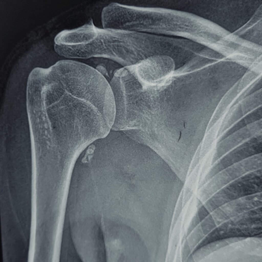
Radiograph of right shoulder (AP)
What are the findings?
- Multiple intra-articular calcified/ossified bodies
- Distribution around the glenohumeral joint and along bicipital groove
- Joint-space preservation
- No pressure erosion/remodelling.
MRI of right shoulder
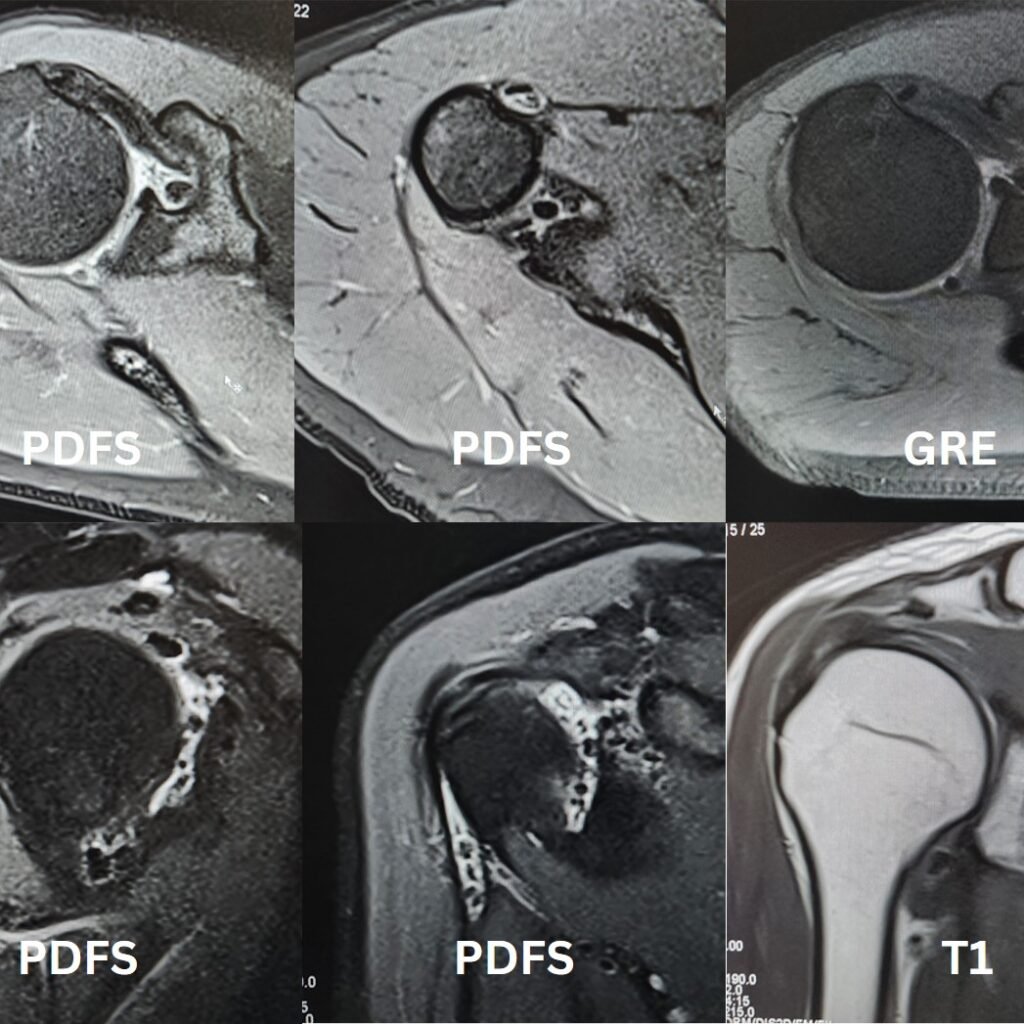
- MRI shows extensive synovial proliferation with multiple relatively similar shape and sized, ossified intra-articular bodies at glenohumeral joint, subscapularis recess, sub coracoid recess and along biceps tendon sheath.
- No degenerative changes seen at glenohumeral Joint.
Differential diagnosis
- Primary synovial chondromatosis
- Secondary synovial chondromatosis
- Synovial osteochondromatosis
- CPPD-related loose bodies
- Osteochondral fragments
IS THIS PRIMARY OR SECONDARY SYNOVIAL CHONDROMATOSIS?
| Feature | Primary | Secondary |
|---|---|---|
| Underlying joint disease | Usually absent | Usually present |
| Number of bodies | Often numerous | Usually fewer |
| Size | Relatively uniform | Variable |
| Shape | Similar morphology | More variable |
| Cartilage/joint degeneration | Usually mild initially | Often prominent |
| Pathogenesis | Metaplastic synovial proliferation | Fragmentation of cartilage/osteophytes |
POINT TO REMEMBER
For a primary case – numerous relatively uniform nodules + synovial involvement + absence of significant pre-existing degenerative joint disease.
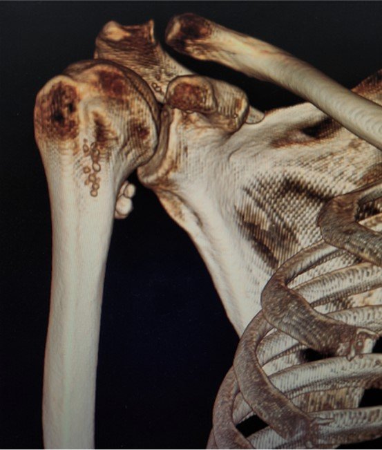
CT- can map entire extent of disease.
LOOK FOR DANGER SIGNS (SYNOVIAL CHONDROSARCOMA)
Malignant transformation is rare, but radiologists should look for:
- Aggressive bone destruction
- Invasion beyond the joint
- Soft-tissue mass
- Increasing lesion size
- Atypical imaging appearance
- Destructive rather than pressure-remodelling changes
LEARNING POINTS
1. Think beyond “loose bodies.”
Numerous similar-sized calcified/ossified bodies should raise suspicion for synovial chondromatosis.
2. Look for the underlying joint.
The absence of significant degenerative disease favors primary rather than secondary disease.
3. Map the entire extent
In the shoulder, evaluate the glenohumeral joint, subacromial/subdeltoid bursa, bicipital groove and other communicating recesses.